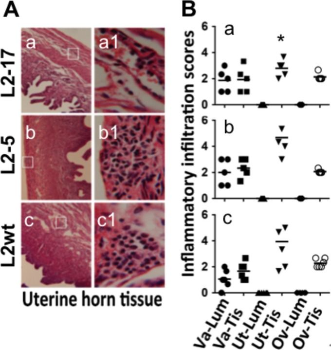
FIG 2.

Effect of CPAF deficiency on inflammatory infiltration in the genital tracts of mice following an intravaginal inoculation. (A) The mice described in the Fig. 1 legend were sacrificed on days 21 to 60 postinfection, and the genital tracts were processed for microscopic detection of inflammatory infiltration. Five mice from each group sacrificed on or close to day 21 were selected for the pathology analysis. The mouse sacrifice dates were matched among the 3 groups. Representative images of uterine horn tissue from each group of mice were taken under 10× (a to c) and 100× (a1 to c1) objective lenses. The areas covered under the 100× lens are marked with white rectangles in the corresponding 10× images. (B) Different sections of the genital tract, including the lumen (Lum) and tissue (Tis) of vagina (Va), uterine/uterine horns (Ut), and oviducts (Ov), were semiquantitatively scored for inflammatory infiltration based on the criteria described in Materials and Methods. The inflammatory scores from each mouse were plotted individually along the y axis. Solid circles stand for the inflammatory scores from Va-Lum, solid squares for scores from Va-Tis, solid standard triangles for scores from Ut-Lum, solid inverted triangles for scores from Ut-Tis, solid diamonds for scores from Ov-Lum, and open circles for scores from Ov-Tis. Note that the inflammatory scores indicated for the uterine/uterine horn tissues (Ut-Tis) of mice intravaginally inoculated with L2-17 (a) are significantly lower (Wilcoxon; *, P < 0.05) than those indicated for mice similarly inoculated with either L2-5 (b) or L2wt (c).
