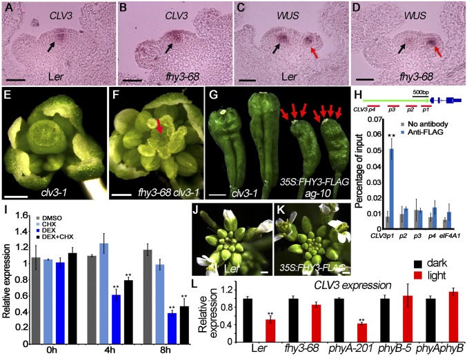
Fig. 4.
CLV3 mediates FHY3 functions in regulating the stem cell pool in the SAM and FM meristem activity. (A–D) In situ hybridization to examine the expression of CLV3 (A and B) and WUS (C and D) in Ler (A and C) and fhy3-68 (B and D). CLV3 signals are marked by a black arrow in A and B. WUS signals are marked by a black arrow in SAM and a red arrow in FM in C and D. (E and F) Flowers of clv3-1 (E) and fhy3-68 clv3-1 (F). Dome-shaped meristem is marked by a red arrow. (G) Representative siliques of clv3-1 (Left) and 35S:FHY3-FLAG ag-10 (Right) plants. Carpels are marked by red arrows. (H) ChIP to measure FHY3 occupancy at CLV3 in 35S:3FLAG-FHY3-3HA fhy3-4 inflorescences. The regions examined are shown on the Upper panel. CLV3 gene structure was shown. (Scale bar, 500 bp.) eIF4A1 served as a negative control. Error bars represent SD from three biological replicates. **P < 0.01 compared with no antibody (negative control). (I) The CLV3 transcript levels in FHY3:FHY3-GR fhy3-4 inflorescences measured by RT-qPCR. (J and K) Inflorescence of Ler (J) and 35S:FHY3-FLAG (K). 35S:FHY3-FLAG developed a larger inflorescence containing more unopened buds than Ler. (L) The CLV3 transcript levels in seedlings of indicated plants after light treatment measured by RT-qPCR. In I and L, UBQ5 served as the internal control. Three biological replicates were performed. Error bars represent SD from three biological repeats. **P < 0.01. (Scale bars: 50 µm in A–D and 500 µm in E–G, J, and K.)

