Fig. 1.
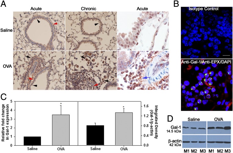
Expression of Gal-1 is induced in allergic lungs. (A) Gal-1 expression in lungs of mice exposed to allergen challenge (or saline alone as control) by IHC. Gal-1 expression in airway epithelial cells (black arrowheads), endothelial cells (red arrow), smooth muscle cells (red arrowheads), inflammatory cells (black arrows), and extracellular spaces (blue arrow) is shown. [Scale bars, 50 µm (Left) and 10 µm (Right).] (B) Lung sections from OVA-exposed mice dual-stained with antibodies against Gal-1 (red) and eosinophil-specific mouse anti-human EPX (green) (Bottom) or rabbit and mouse IgG as controls (Top). (Scale bar, 10 µm.) (C) Gal-1 mRNA (Left) and protein (Right) expression in lungs of OVA (acute)-challenged or saline-exposed mice. (D) Gal-1 expression in lung tissue of three representative mice (M1–M3) for each group. Data are representative of n = 6 mice per group in A and n = 3 mice per group in B. Combined data (mean ± SEM) of n = 5–7 mice per group are shown in C. *P < 0.05 (Left) and *P < 0.01 (Right) in C for control versus allergen-challenged group.

