Fig. 2.
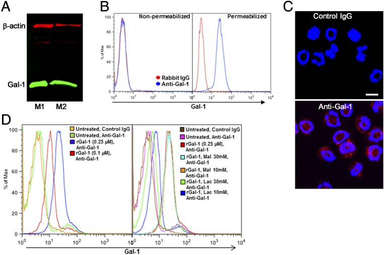
Murine eosinophils express Gal-1 intracellularly and bind to Gal-1 on the cell surface. (A) Expression of Gal-1 in lysates of BM eosinophils from two representative mice (M1 and M2) by Western blot analysis. (B) Expression of Gal-1 in BM-derived eosinophils by flow cytometry. Note that Gal-1 expression is detected only in permeabilized cells. (C) IF staining of permeabilized eosinophils with anti–Gal-1 (Bottom) or rabbit IgG (Top). (Scale bar, 10 µm.) (D) Dose-dependent Gal-1 binding to nonpermeabilized eosinophils in the absence (Left) or presence (Right) of increasing concentrations of lactose (Lac) or maltose (Mal). Data are representative of three independent experiments with eosinophils from different mice.

