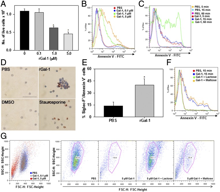
Fig. 4.
Exogenous Gal-1 induces eosinophil apoptosis. (A) Effect of rGal-1 on the absolute number of live cells by trypan blue dye exclusion at the indicated doses. (B and C) Flow cytometric analysis after annexin V staining of eosinophils exposed to different concentrations of rGal-1 and eosinophils treated with 5.0 µM rGal-1 for different durations of time, respectively. (D) Analysis of cell death in eosinophils (stained brown) after rGal-1 or staurosporine (positive control) treatment by TUNEL assay. (Scale bar, 50 μM.) (E) Annexin V staining of peripheral blood leukocytes exposed to rGal-1. Annexin V positivity of cells gated for Siglec-F–positive staining (eosinophils) is shown. (F) Inhibition of Gal-1–induced apoptosis by lactose but not maltose (negative control). (G) Effect of rGal-1 on eosinophil shape change. FSC versus side scatter dot plots are shown. (G, Left) Dose-dependent cell shrinkage in Gal-1–treated eosinophils. (G, Right) Blockade of this effect by lactose but not maltose. Combined data (mean ± SEM) of three independent experiments with cells from n = 3–7 mice are shown in A and E. Data shown in B–D, F, and G are representative of two or three experiments with eosinophils from different mice. *P < 0.01 in A and E for comparison with PBS-treated cells.

