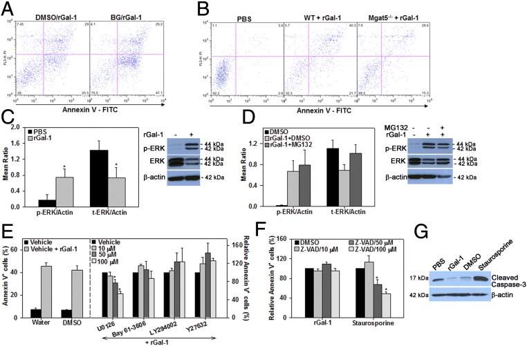
Fig. 5.
Gal-1–induced apoptosis is mediated by complex branched N-glycans and involves activation of the ERK(1/2) signaling pathway. (A) Involvement of O-glycans in Gal-1–induced apoptosis. Eosinophils cultured in medium containing BG or vehicle (DMSO) were treated with rGal-1 (5 µM) and examined for annexin V positivity. (B) Requirement of complex branched N-glycans for Gal-1–induced apoptosis. WT and Mgat5−/− eosinophils were treated with rGal-1 and examined for annexin V positivity. (C) Induction of ERK(1/2) phosphorylation in Gal-1–treated eosinophils determined by densitometric analysis of Western blots. (D) Inhibition of ERK(1/2) degradation by proteasome inhibitor MG132 determined by densitometric analysis of Western blots. (E) Inhibition of Gal-1–induced apoptosis by MEK1/2 inhibitor. (E, Right) Annexin V staining of eosinophils pretreated with the indicated inhibitors of signaling molecules and then with rGal-1 relative to vehicle alone is shown. (E, Left) Annexin V positivity in vehicle-treated versus rGal-1–treated cells is shown. (F) Caspase-independent apoptosis of eosinophils by Gal-1. Cells were pretreated with caspase inhibitor (pan; Z-VAD-FMK) at the indicated doses (or vehicle) followed by rGal-1 or staurosporine and examined for annexin V positivity. (G) Western blot of Gal-1–treated eosinophils with antibodies against cleaved caspase-3. Data shown in A and B are representative of three experiments and in G of two experiments with eosinophils from different mice. Combined data (mean ± SEM) of three or four independent experiments in C and D and four to six independent experiments in E and F are shown. *P < 0.05 in C, *P < 0.05 for 50 µM and *P < 0.01 for 100 µM U0126 in E, and *P < 0.01 for 50 and 100 µM Z-VAD in F for comparison with PBS or vehicle-treated cells.

