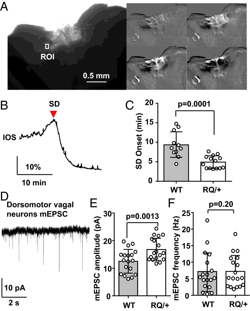
Fig. 8.
Lowered SD threshold in dorsal vagal complex of the RQ mutant brainstem. (A–C) Characterization of SD generated in dorsal medulla in vitro. Brainstem slices containing the dorsal vagal complex were exposed to OGD solution (0% O2, 5 mM glucose), and SD was detected by monitoring intrinsic light transmission signals in the region of interest (ROI) positioned in the lateral NTS (A and B). A raw image of an acute brainstem slice (Left large) and a sequence of ratio images showing spreading light transmission changes following OGD exposure (Right). The latency to SD onset was significantly shorter in the RQ mutant (C). WT, 11 slices from 4 mice; RQ, 15 slices from 5 mice. (D–F) Characterization of mEPSC in dorsomotor vagal (DMV) neurons. (D) Representative trace of mEPSCs recorded from a DMV neuron. The mean amplitude was significantly larger in RQ mutant neurons (E), whereas the mean frequency did not differ (F). WT, 18 neurons from 4 mice; RQ, 18 neurons from 4 mice. Bar graph, mean ± standard deviation.

