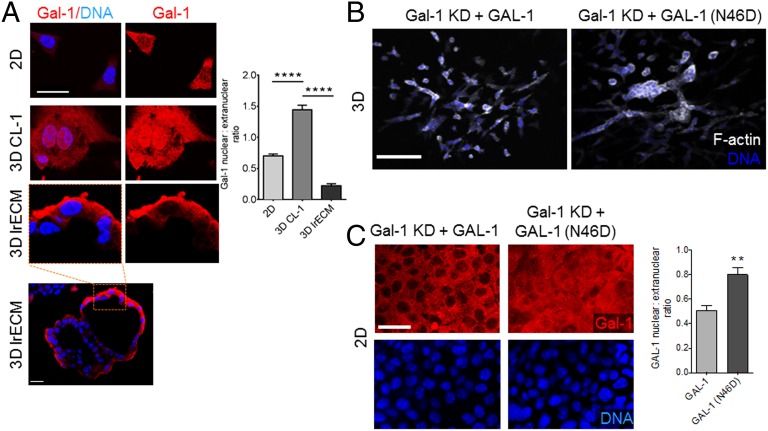
Fig. 3.
Glycan recognition by Gal-1 is dispensable for epithelial branching. (A) Immunofluorescence micrographs of EpH4 cells cultured in 2D (Top), on top of 3D CL-I gel (Middle), and on top of 3D lrECM gel (Bottom Left: acinar-like architecture with lumen. Scale bar, 20 µm) and stained for Gal-1 (depicted in red) and DNA (depicted in blue). Quantification of Gal-1 nuclear:extranuclear ratio for EpH4 cells cultured in 2D and 3D conditions. (Scale bar, 25 µm.) (B and C) Fluorescence micrographs of Gal-1 KD EpH4 cells ectopically expressing GAL-1 (Left) or GAL-1 (N46D) (Right) fusion proteins in 3D (Top) or 2D (Bottom) and stained for F-actin (depicted in white) and DNA (depicted in blue). Cells expressing GAL-1 (N46D), a mutant with attenuated glycan binding, invade and branch when cultured in 3D. Quantification of GAL-1 nuclear:extranuclear ratio shows GAL-1 (N46D) is concentrated in the nucleus. (Scale bar, 100 µm.) For all bar graphs, error bars represent S.E.M. Statistical significance is given by **P < 0.01; ****P < 0.0001.

