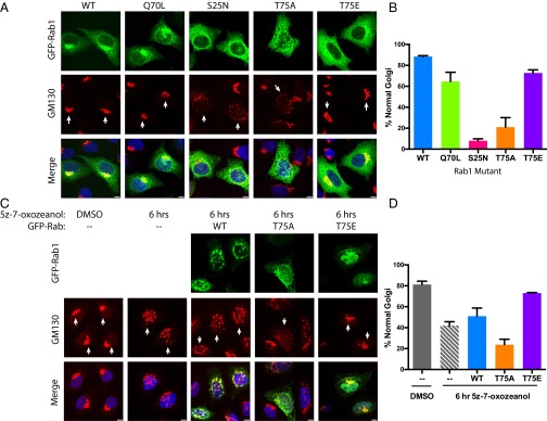
Fig. 5.
Fragmentation of the Golgi is a result of overexpression of Rab1T75A or inhibition of TAK1. (A) Representative images of HeLa cells transfected with GFP-Rab1 in green and stained for GM130, a cis-Golgi marker in red, and DAPI in blue. (Scale bar, 5 μm.) (B) Quantitation of immunofluorescence experiment (n = 3 replicates, 33 cells per replicate). (C) Representative images of HeLa cells stained for GM130 after dosing with 2.5-μM TAK1 inhibitor 5z-7-oxozeaenol for 6 h and preceding transfection with GFP-Rab1 where indicated. (Scale bar, 5 μm.) (D) Quantitation of immunofluorescence experiment (n = 3 replicates, 33 cells per replicate).

