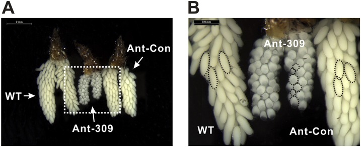
Fig. S2.
Phenotypic analysis of ovaries dissected from WT, Ant-309–injected, and Ant-Con–injected female mosquitoes at the 36-h PBM with scale bars of 2 mm (A) and 0.9 mm (B), respectively. The dashed circles indicate the follicle shape and size. The nurse cells were visible in the Ant-309–treated ovary.

