Fig. 2.
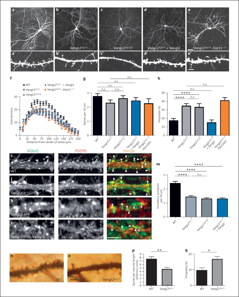
Differentiation phenotypes in Vangl2Lp forebrain neurons. a-e EGFP-transfected cultured neurons from WT (a), Vangl2Lp/+ (b), Vangl2Lp/Lp (c), Vangl2Lp/+ + Vangl2 (d) and Vangl2Lp/+; Dact1-/- (e). a'-e' Corresponding dendritic segments at a higher magnification. f-h Quantification. Vangl2Lp neurons have simpler dendritic arbors (f), no reduction in total density of dendritic projections (spines + filopodia) (g), but a larger proportion of immature (filopodial) dendritic projections (h) than controls; only the last phenotype is rescued by recombinant expression of Vangl2 (blue bar; color refers to the online version only). i-l Immunostaining for glutamatergic synapse markers along dendrite segments from WT (i), Vangl2Lp/+ (j), Vangl2Lp/Lp (k) and Vangl2Lp/++ Vangl2 (l) cultured neurons. m Quantification. n, o Segments of apical dendrite from a Golgi-stained pyramidal neuron in hippocampal CA1 of WT (n) and Vangl2Lp/+ (o) littermates. Quantification of total spines (p) and immature (filopodial) spines (q). Scale bars = 30 μm (a-e); 5 μm (a'-e', i-l, n, o). n.s. = p > 0.05, * p ≤ 0.05; ** p ≤ 0.01, **** p ≤ 0.0001.

