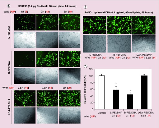
Figure 7. . Comparison of gene delivery efficiency and cytotoxicity of L-PEI (25 kDa), B-PEI (25 kDa) and LGA-PEI polymer in HEK293 and PANC-1 cells.
(A) Delivering GFP containing plasmid to HEK293 cells. Cells were seeded onto the 96-well plate overnight. Three concentrations of delivery agents (1:1, 2:1 and 3:1 w/w for L-PEI and B-PEI; 2.5:1, 3.5:1 and 5:1 w/w for LGA-PEI polymer) and 0.2 μg GFP plasmid DNA/well were used for transfection for 24 h. Green florescence signal as transfection efficiency was examined under a fluorescence microscope, and cellular density as potential cytotoxicity was evaluated with phase contrast microscopy analysis. (B) Delivering GFP containing plasmid to PANC-1 cells. L-PEI (2:1 w/w), B-PEI (2:1 w/w) or LGA-PEI (3.5:1 w/w) and 0.2 μg GFP plasmid DNA/well were used for transfection in the 96-well plate for 48 h. Green florescence signal and cellular density were recorded. (C) Cell viability after gene delivering in PANC-1 cells. L-PEI (2:1 w/w), B-PEI (2:1 w/w) or LGA-PEI (3.5:1 w/w) and 0.2 μg GFP plasmid DNA/well were used for transfection in the 96-well plate for 48 h. Cell viability was determined by MTT assay.
*p < 0.05, n = 6.
LGA-PEI: Lactic-co-glycolic acid-modified polyethylenimine; PEI: Polyethylenimine.

