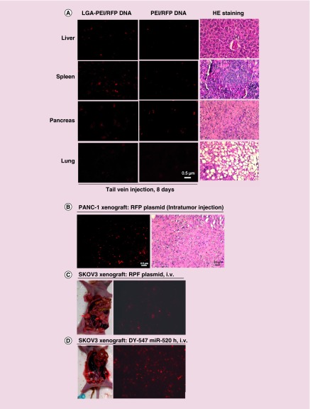
Figure 8. . Delivery efficiency of LGA-PEI polymer/nucleic acids into mouse models.
LGA-PEI (0.5:1 w/w) polymer was used. (A) Organ distribution. Plasmid DNA (30 μg), with either LGA-PEI polymer or PEI, was injected via tail vein into mice three-times during 5 days; mice were sacrificed on day 8 (n = 3). Fluorescence microscope imaging was performed for liver, spleen, pancreas and lungs. H&E staining for these organs was performed for observation of the tissue structure, which is normal. (B) Pancreatic xenograft tumor in a nude mouse model. Nude mice (n = 3) were injected subcutaneously with pancreatic cancer cell line AsPC-1 for 2 weeks. LGA-PEI/RFP DNA (10 μg) NPs were directly injected into the tumor. Three days later, the tumors were harvested and the red fluorescence analyzed. H&E staining showed xenograft cellular morphology. (C) Human ovarian cancer cell line (SKOV3) was orthotopically injected into the bilateral ovaries of female nude mice (n = 5). When xenograft tumors developed in about 5 weeks, LGA-PEI/RFP DNA (30 μg) NPs were injected via tail vein into the nude mice three-times in 5 days. Five days later, the tumors were harvested and the red fluorescence analyzed. (D) Orthotopic SKOV3 tumors in nude mice (n = 3) were established in 5 weeks, then LGA-PEI/miR-520h mimic (DY-547 tag, 15 μg) NPs were injected into the mouse via tail vein once. Five days later, the tumors were harvested and the red fluorescence was analyzed.
LGA-PEI: Lactic-co-glycolic acid-modified polyethylenimine; PEI: Polyethylenimine.

