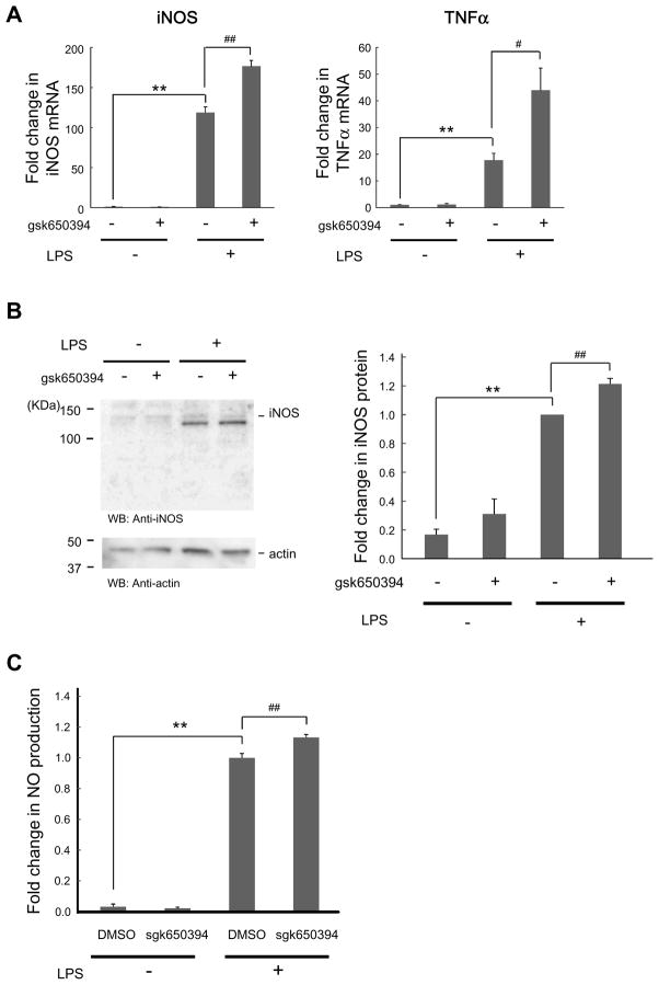
Figure 3. SGK inhibition increases LPS-induced inflammatory responses.
Cells were incubated in the absence or presence of gsk650394 (1 μM) for 30 min, followed by LPS (500 ng/ml) administration for 4 h (A), 8 h (B), and 24 h (C). (A) After RNA extraction and reverse transcription to synthesize cDNA, quantitative real-time PCR was performed to monitor mRNA levels of iNOS and TNFα in cells treated with indicated reagents. n = 3. (B) BV-2 cell lysates were analyzed by immunoblotting with the indicated antibodies. n = 4. (C) Media were taken and NO was measured by assessing nitrite, a natural metabolic product of NO. n = 6–8. ** p < 0.01 vs LPS, # p<0.05, ## p < 0.01 vs DMSO (0 μM gsk650394), Student’s t-test.

