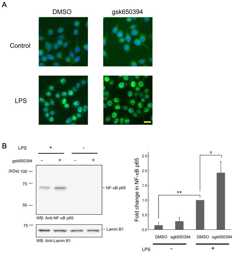
Figure 4. SGK inhibition enhances NF-κB signaling activity.
(A) BV-2 cells were incubated in the absence or presence of gsk650394 (1 μM) for 30 min, followed by LPS (500 ng/ml) administration for 30 min, and then fixed. NF-κB p65 protein was visualized by indirect immunofluorescence staining using an antibody for NF-κB p65. For nuclear staining, the cells were stained with DAPI. Bar = 20 μm. (B) Cells were treated as described in (A) and nuclear samples were extracted, followed by Western blotting using anti- NF-κB p65 antibody. Protein loading was monitored by anti-lamin B1 antibody. n = 4. ** p < 0.01 vs LPS, # p<0.05 vs DMSO, Student’s t-test.

