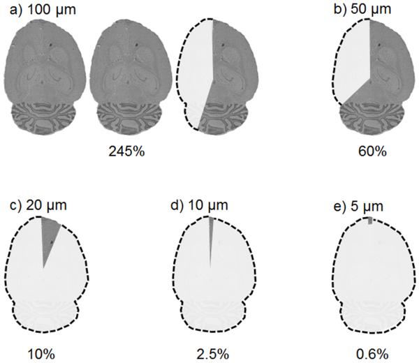
Figure 2.
A depiction of an IMS experiment showing that, given a defined circular tissue area (d=1.5 cm, represented by one cartoon brain image, images not shown to scale) and a 1 pixel/second acquisition speed, the amount of tissue that can be sampled in 12 hours is dependent upon the spatial resolution of the experiment. Percentages indicate the approximate proportion of one brain section that can be measured at the indicated spatial resolution.

