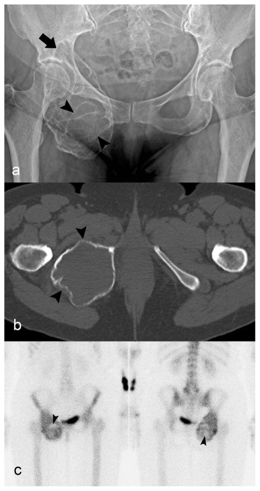
Fig. 1.
Giant cell tumor of bone of the ischium. a) Radiograph of pelvis shows an expansile lytic lesion causing marked cortical thinning. The rim is partially sclerotic (arrow) and there are septations (arrowheads) in the lesion. b) Axial unenhanced CT image demonstrates marked cortical thinning (arrowhead) without periosteal reaction or mineralized matrix. c) Bone scan shows the radiotracer uptake predominantly at the periphery and central photopenia (arrowheads)

