Fig. 1.
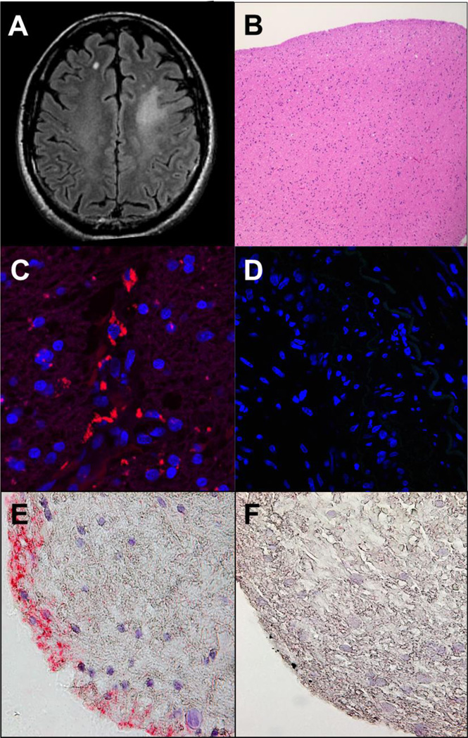
Brain imaging and histology, and immunostaining of brain biopsy for varicella zoster virus antigen. Axial FLAIR brain MRI demonstrates a focal area of T2 hyperintensity in the left anterior centrum semiovale with ill-defined margins, without mass effect or volume loss (A). No post-contrast enhancement was seen (images not shown). Hematoxylin and eosin stained paraffin-embedded section of the brain biopsy shows well-fixed minimally hypercellular white matter without evidence of neoplasm, inflammation, vasculitis or necrosis (B). These findings were confirmed with special immunostains for GFAP, CD68 and IDH1 mutation (data not shown). Indirect immunofluorescence with mouse monoclonal anti-VZV antibodies directed against both the VZV immediate early (IE) 62 and the late VZV gE proteins applied to the brain biopsy, as described by Halling et al. [1], revealed VZV antigen (red) in the cytoplasm of brain cells (C), but not when primary antibody was omitted (D); the same anti-VZV antibody did not stain normal brain tissue [1]. VZV antigen was also detected by immunohistochemistry with rabbit monospecific polyclonal anti-VZV IE 63 antibody (E, pink color), as described by Gilden et al. [2], in a different section of the same brain biopsy, but not in another section of the same brain biopsy when normal rabbit serum was substituted for primary rabbit anti-VZV IE 63 antibody on (F). Magnification: 100X (B); 630X (C and D); 600X (E and F).

