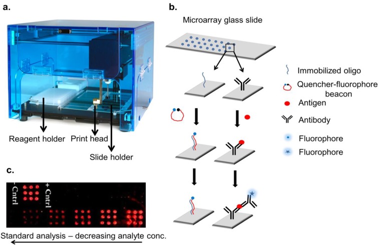
Figure 1.
Illustration of microarray technology. An array printer is depicted in (a) which has a dedicated space for holding reagent plates and slides. An automated syringe cum pin is filled up with specific reagent followed by spotting in specific spot-sizes and electronically controlled volumes (Table 2); (b) Methodology of creating oligo/antibody array is depicted. Each spot on the slide holds immobilized oligonucleotides or antibodies which were later used for analyte detection; (c) An assay in a microarray format is shown with immobilized anti-fetuin antibody detecting various concentrations of fetuin and detection performed with Cy5 anti-fetuin antibody. Spot size of the array is 2 mm each, with a spotting volume of 1 µL anti-fetuin capture antibody.

