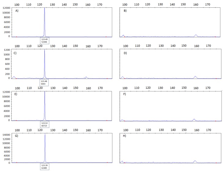
Figure 3.
Electropherograms used to evaluate the presence of mycobacterium in donor blocks from a previously confirmed positive and negative tissue sample. (A) positive control and (B) negative control. Tissue samples were alternated in the tissue arrayer three times leading to the following results: (C,E,G) mycobacterium tuberculosis samples and (D,F,H) mycobacterium tuberculosis negative samples. No evidence for cross-contamination between samples.

