Figure 2.

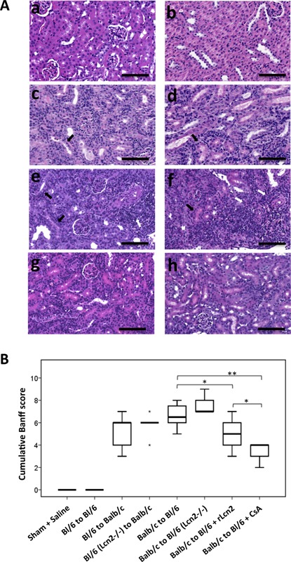
Effect of lipocalin 2 (Lcn2) on histopathology of the mouse allografts. (A) Representative images of hematoxylin and eosin–stained sections of the kidneys from sham‐operated mice (a), C57Bl/6 (Bl/6) kidney isografts (b) and the kidney allografts Bl/6 to Balb/c (c), Bl/6 Lcn2−/− to Balb/c (d), Balb/c to Bl/6 (e), Balb/c to Bl/6 Lcn2−/− (f), Balb/c to Bl/6 (treated with recombinant Lcn2:Sid:Fe complex (rLcn2; 250 μg, perioperatively) (g), Balb/c to Bl/6 (treated with cyclosporine A [10 mg/kg body weight, daily]) (h), harvested at posttransplant day 7 are shown. Black and white arrows highlight the examples of tubulitis and periarterial lymphocytic aggregates, respectively. (B) The cumulative Banff score of the prominent histopathological lesions of the kidney allografts is presented by a box plot (n = 6, except the groups Balb/c to Bl/6 and Balb/c to Bl/6 + rLcn2 n = 8). Scale bars = 100 μm. *p < 0.05, **p < 0.01.
