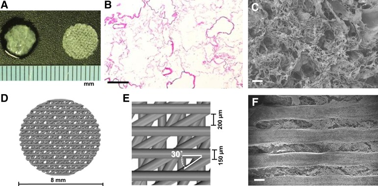
Figure 1.
Matrix architecture of three-dimensional extracellular matrix (ECM) and bioplotted poly-l-lactic acid (PLLA)-collagen scaffolds. (A): ECM scaffold (left) and porous PLLA-collagen scaffold (right). (B, C): Surface view of the ECM scaffold by hematoxylin and eosin staining (B) and scanning electron microscopy (SEM) imaging (C). (D, E): Computer-aided schematic diagram of the PLLA-collagen scaffold showing struts and pores. (F): SEM micrograph of the surface of PLLA-collagen scaffold depicting type 1 collagen infused between PLLA struts. Scale bars = 200 µm.

