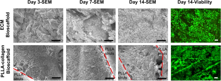
Figure 2.
Induced pluripotent stem cell (iPSC) hepatocytes survive and grow within ECM and PLLA-collagen scaffolds. SEM micrographs of recellularized ECM and PLLA-collagen scaffolds with iPSC hepatocytes over 14 days during an in vitro time course study. Red dashed lines delineate boundaries between PLLA bioprinted struts and infused type 1 collagen within three-dimensional bioprinted PLLA-collagen scaffolds. Staining of iPSC hepatocytes within ECM scaffolds or PLLA-collagen scaffolds showed live cells (green) with minimal dead cells (red) at day 14. Scale bars = 50 µm. Abbreviations: ECM, extracellular matrix; PLLA, poly-l-lactic acid; SEM, scanning electron microscopy.

