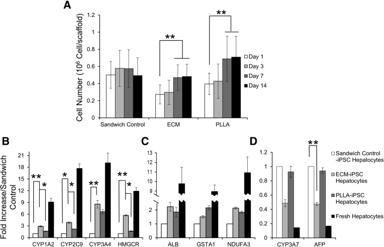
Figure 4.
iPSC hepatocyte culture within three-dimensional scaffolds leads to increased proliferation and expression of mature liver-specific markers. (A): Cell proliferation of iPSC hepatocytes in ECM scaffolds, PLLA-collagen scaffolds, and sandwich controls (n = 4 samples for each group). Quantitative reverse transcription-polymerase chain reaction of metabolic and synthetic genes in iPSC hepatocytes that either changed (B) or remained stable (C) in response to growth in ECM and PLLA-collagen scaffolds compared with growth in standard sandwich control culture at day 14. Fresh, human hepatocytes are shown for comparison. (D): Expression of fetal genes CYP3A7 and AFP in iPSC hepatocytes in ECM scaffolds or PLLA-collagen scaffolds compared with control sandwich culture at day 14 (∗, p < .05; ∗∗, p < .01; n = 4 for each group). Abbreviations: AFP, α-fetoprotein; ECM, extracellular matrix; HMGCR, human 3-hydroxy-3-methylglutaryl-coenzyme A reductase; iPSC, induced pluripotent stem cell; PLLA, poly-l-lactic acid.

