Figure 3.
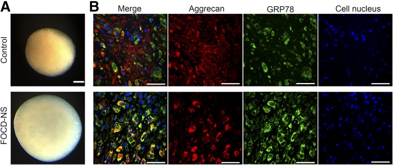
Morphologic and immunohistochemical analysis of micromass cultures derived from control and FOCD-NS BM-MSCs. (A): Images showing the morphology of day-35 micromass cultures. Scale bar, 200 µm. (B): Confocal microscopy images showing double staining of aggrecan (red), GRP78 (green), and overlapping areas (yellow) on day-35 cultures. Cell nuclei stained with DAPI (blue). Scale bar, 50 µm. Abbreviations: BM-MSC, bone marrow mesenchymal stem cell; DAPI, 4′,6-diamidino-2-phenylindole; FOCD-NS, familial osteochondritis dissecans from northern Sweden.

