Figure 6.
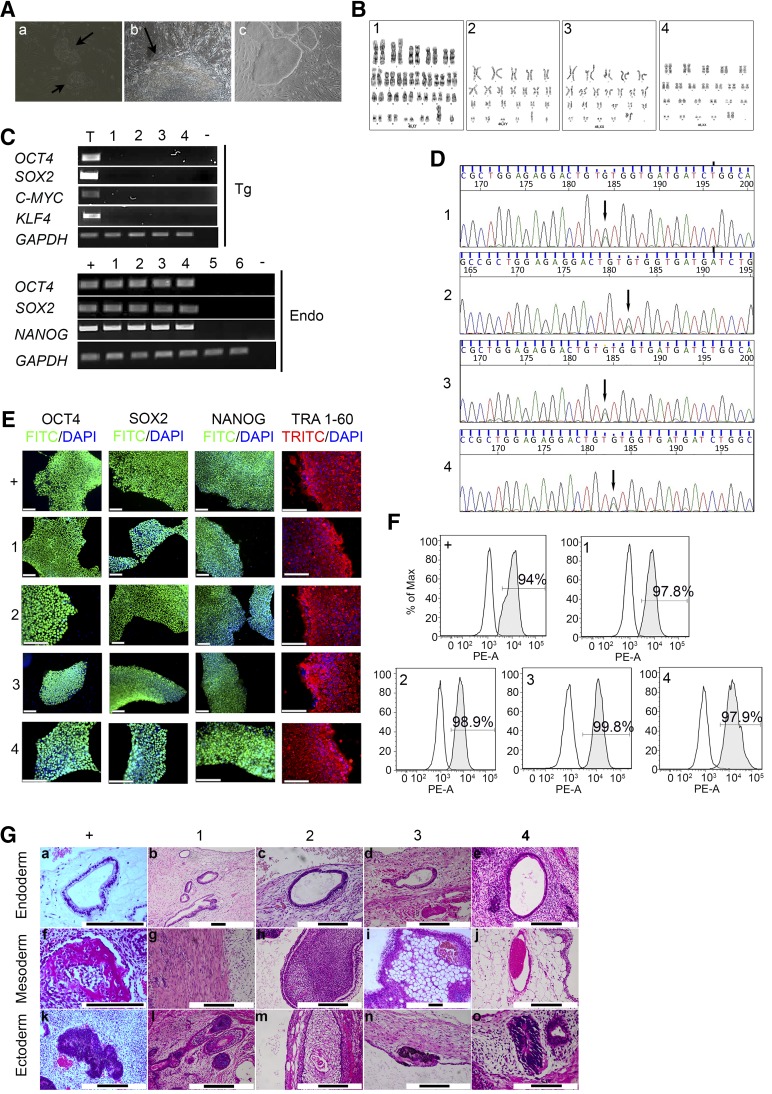
Generation and characterization of iPSCs: FOCD-NS1-iPSC-2 (1), FOCD-NS1-iPSC-30 (2), FOCD-NS2-iPSC-9 (3), and FOCD-NS2-iPSC-13 (4). Normal iPSC line 33D-6 was the positive control (+). Skin fibroblasts obtained from FOCD-NS1 (5) and FOCD-NS2 (6) were included. (A): Reprogrammed fibroblasts underwent mesenchymal–epithelial transition at day 6 (a) and formed an early iPSC-like colony at day 14 (b) (arrows; magnification ×20). Morphology of iPSC colonies (magnification ×10) (c). (B): G-banding analysis showed normal karyotype in FOCD-NS iPSCs. (C): RT-PCR data showing expression of retroviral transgenes (Tg) and endogenous pluripotent genes (Endo) in FOCD-NS iPSCs. Nontemplate controls (−) were included in each PCR reaction. RNA extracted from retroviral-infected 293T cells acted as positive controls for detection of Tg factors (T). (D): DNA sequencing confirming the aggrecan mutation in FOCD-NS iPSCs (arrows). (E): Expression of pluripotency markers in FOCD-NS iPSCs by immunostaining. Scale bar, 120 µm. (F): Flow cytometry analysis of the expression of SSEA4. White histograms represent isotype controls, and gray overlays represent SSEA4; percentage positive cells shown within histograms. (G): Histologic analysis of hematoxylin and eosin-stained teratoma sections derived from iPSCs. Detected tissue types included endoderm (top row), mesoderm (middle row), and ectoderm (bottom row). Black scale bar, 200 µm. Abbreviations: DAPI, 4′,6-diamidino-2-phenylindole; Endo, endogenous pluripotent genes; FITC, fluorescein isothiocyanate; FOCD-NS, familial osteochondritis dissecans from northern Sweden; GAPDH, glyceraldehyde-3-phosphate dehydrogenase; iPSC, induced pluripotent stem cell; PE-A: phycoerythrin-labeled antibody; RT-PCR, reverse-transcription polymerase chain reaction; SSEA, stage-specific embryonic antigen; Tg, retroviral transgenes; TRITC, tetramethylrhodamine isothiocyanate.

