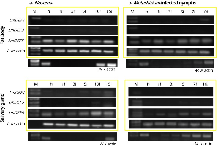
Fig 5. Tissue specificity and developmental expression patterns of LmDEFs.
a, spatiotemporal expression of LmDEFs in N. locustae infected fat body and salivary gland cells. b, same, but with M. anisopliae. M, DNA ladder; h, healthy insect; 1i, 3i, 5i,7i, 10i, and 15i the RNA of tissues collected on the 1st, 3rd, 5th, 7th, 10th, and 15th days after inoculation with pathogens; L.m. actin, locust actin gene. N.l., Nosema spores and M.a, Metarhizium hyphae as positive controls with RT-PCR. The locust actin gene was used as a control for the integrity of the cDNA templates. Amplification products were analyzed on agarose gels and visualized by UV illumination after ethidium bromide staining. All tissues were dissected from gregarious locusts.

