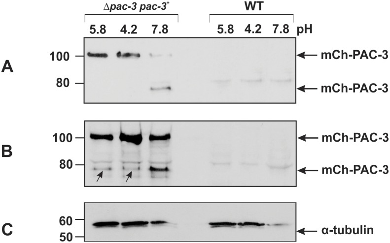
Fig 3. PAC-3 proteolytic processing at alkaline pH.
PAC-3 protein levels were detected by western blot using the polyclonal anti-mCherry antibody and cell extracts from the wild-type and complemented (Δpac-3 pac-3+) strains cultivated at pH 5.8 at 30°C for 24 h and then shifted to pH 4.2 and 7.8 at 30°C for 1 h. (A) Aliquots of 50 μg of total protein were loaded onto the gel. (B) Aliquots of 70 μg of total protein were loaded onto the gel. The arrows indicate the processed mCh-PAC-3 form at pH 5.8 and 4.2. (C) The protein α-tubulin was used as the loading control. The plots represent one of the three independent experiments. The numbers on the left represent the molecular weight in kD.

