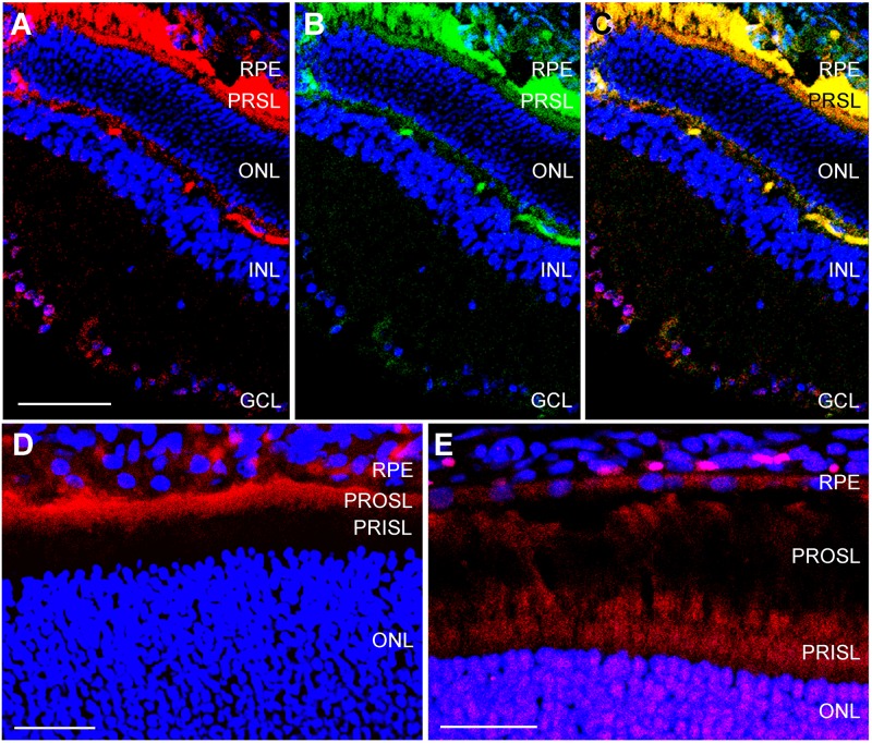
Fig 4. Confocal microscopy images of adult retina.
Representative confocal microscopy images of the retina of adult rats exposed for 3 h to hypothermia and then for 24 h at room temperature. Sections were stained with antibodies against CIRP (red, A,D) and RBM3 (green in B, red in E). To test for colocalizations, C represents an overlay of A and B. RPE = retinal pigment epithelium, PRSL = photoreceptor segment layer, ONL = outer nuclear layer, INL = inner nuclear layer, GCL = ganglion cell layer,. Bar for A-C = 50 μm. Bar for D = 25 μm. Bar for E = 20 μm.

