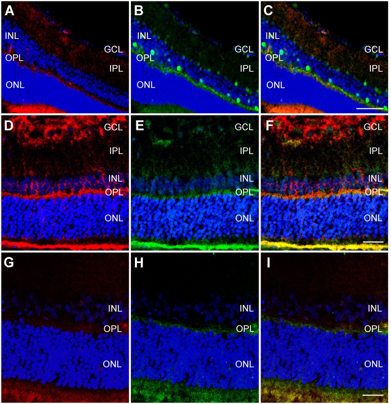
Fig 7. Colocalization of retinal markers with RBM3 in adult retina.
Representative confocal microscopy images of colocalizations in hypothermic adult rat retina between RBM3 (A,E,G) and cell specific markers calbindin (B), glutamine synthetase (D), and recoverin (H). The third column is a combination of the first two; a yellow hue represents colocalization. GCL = ganglion cell layer, IPL = inner plexiform layer, INL = inner nuclear layer, OPL = outer plexiform layer, ONL = outer nuclear layer. Bar for A-C = 50 μm. Bar for D-F = 25 μm. Bar for G-I = 25 μm.

