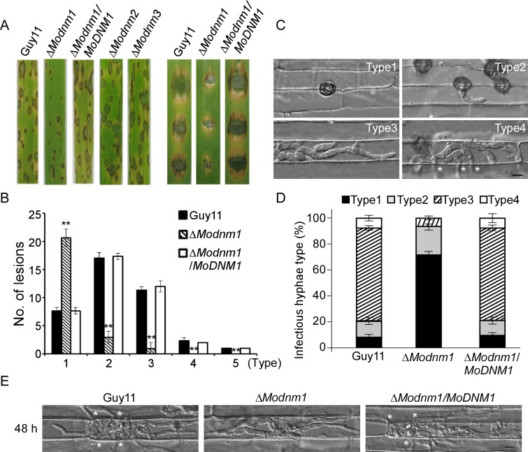
Fig 2. MoDnm1 is important for full virulence of M. oryzae.
(A) Rice (Oryza sativa cv. CO39) seedlings (left) were sprayed with conidial suspensions and examined 7 dpi. Detached barley (Hordeum vulgare cv. Four-arris) (right) leaves were drop-inoculated with conidial suspensions and examined 5 dpi. (B) Quantification of lesion types (0, no lesion; 1, pinhead-sized brown specks; 2, 1.5 mm brown spots; 3, 2–3 mm grey spots with brown margins; 4, many elliptical grey spots longer than 3 mm; 5, coalesced lesions infecting 50% or more of the leaf area). Lesions were measured at 7 dpi, numbers within an area of 1.5 cm2 were counted, and experiments were repeated three times with similar results. Asterisks represent significant differences (Duncan's new multiple range test, p<0.01). (C, D) Detailed observation and statistical analysis of invasive growth in rice sheath cells at 36 hpi. For each sample, appressorium penetration sites (n = 100) were checked and the invasive hyphae (IH) were rated from type 1 to 4. Error bars represent ±SD from three independent experiments. Bar = 5 μm. Asterisks indicate IH extended to surrounding cells. (E) Comparative analysis of invasive hyphal growth of the wild type, ΔModnm1 mutant and complemented strains in rice sheath cells at 48 hpi. Asterisks indicate IH extended to surrounding cells.

