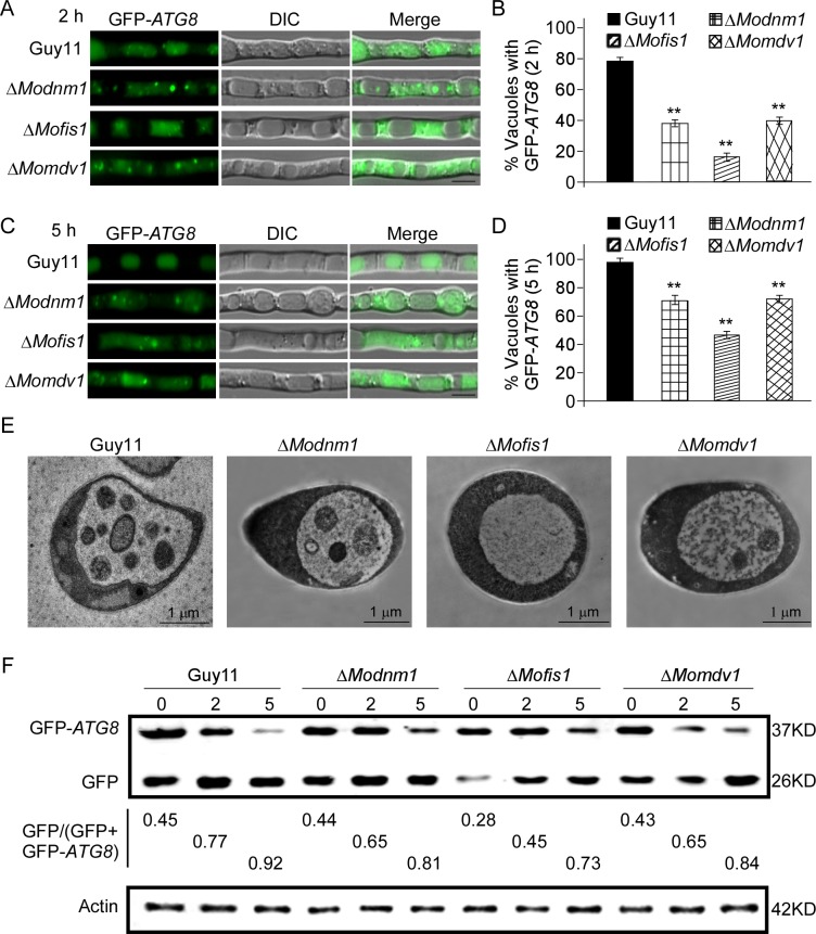
Fig 13. MoDnm1, MoMdv1 and MoFis1 are involved in autophagy.
(A and C) The wild type, ΔModnm1, ΔMofis1, and ΔMomdv1 strains transformed with GFP-Atg8 were cultured in MM-N (nitrogen starvation minimal medium) for 2 h or 5 h, and the autophagy activity was observed by Axio Observer A1 Zeiss inverted microscope. (B and D) Autophagy activity was assessed by means of translocation of GFP-Atg8 into vacuoles (n = 100). Bars with asterisks represent significant differences (Duncan's new multiple range method p<0.01). Bar = 5μm. (E) Transmission electron microscopy (Hitachi H-7650) observation of hyphae cultured in nitrogen starvation MM–N medium for 5 h. Bar = 1 μm. (F) Immunoblotting was performed with anti-GFP and anti-β-Actin antibodies. The extent of autophagy was estimated by calculating the amount of free GFP compared with the total amount of intact GFP-Atg8 and free GFP (the numbers underneath the blot). Densitometric analysis was performed by Image-pro plus (Media Cybernetics Inc., Shanghai, China).

