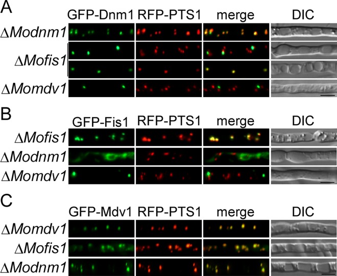
Fig 15. MoMdv1 contributes to the peroxisomal localization of MoDnm1 and MoFis1.
(A) GFP-Dnm1 and RFP-PTS1 were co-expressed in ΔModnm1 (top), ΔMofis1 (middle), and ΔMomdv1 mutants (bottom). Punctate GFP-Dnm1 localization was observed in hyphae by Axio Observer A1 Zeiss inverted microscope. PTS1, a peroxisome marker protein. Bar = 5 μm. (B) GFP-Fis1 and RFP-PTS1 were co-expressed in ΔMofis1 (top), ΔModnm1 (middle), and ΔMomdv1 mutants (bottom). GFP-Fis1 localization was observed in hyphae by Axio Observer A1 Zeiss inverted microscope. PTS1, a peroxisome marker protein. Bar = 5 μm. (C) GFP-Mdv1 and RFP-PTS1 were co-expressed in ΔMomdv1 (top), ΔMofis1 (middle), and ΔModnm1 mutants (bottom). GFP-Mdv1 localization was observed in hyphae by Axio Observer A1 Zeiss inverted microscope. PTS1, a peroxisome marker protein. Bar = 5 μm.

