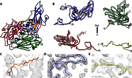
Fig. 3. Rigid body movements of virus proteins to form the CVB3 A-particle.

(A) The structure of the CVB3 A-particle is shown as a ribbon diagram with VP1, VP2, VP3, and VP4 colored according to convention (blue, green, red, and yellow, respectively), with pocket factor rendered as an orange surface. (B) The four structural proteins of the A-particle (blue, green, red, and yellow) are aligned with the structure of CVB3 (dark gray) to illustrate the outward movements. (C to E) The structures of the pocket factor (orange wire), the N terminus of VP1 (blue wire), and the VP4 of CVB3 are shown fitted into the corresponding A-particle densities.
