
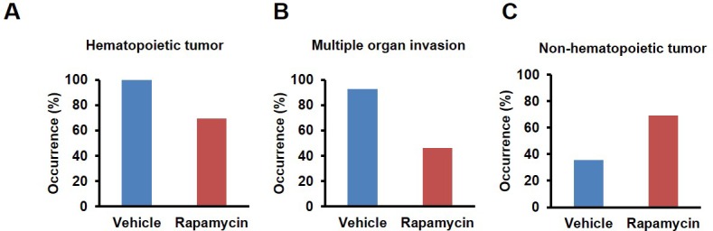
Figure 3. Rapamycin injection at 8 mg/kg/day for 3 months alters cancer incidence of female mice.
(A) Hematoxylin and eosin (H&E) sections of multisystemic aggressive lymphoma (top) and atypical plasmacytoid lymphoma (bottom) from rapamycin-treated female mice. Arrows indicate a bizarre mitotic figure (top) and round cells with strongly eosinophilic cytoplasm (plasmacytoid morphology, bottom). Original magnification 60x. Bar = 10 µm. (B) Hematopoietic cancer incidence of rapamycin-treated (16 female) and vehicle-treated (12 female) mice. (C) Incidence of multiple organ invasion of hematopoietic tumors in rapamycin-treated (16 female) and vehicle-treated (6 female) hematopoietic tumor-bearing mice. (D), Non-hematopoietic cancer incidence of rapamycin-treated (16 female) and vehicle-treated (12 female) mice. *p<0.05. **p<0.01.

Figure 3—figure supplement 1. Morphologies of aggressive hematopoietic tumors observed in rapamycin injected females.
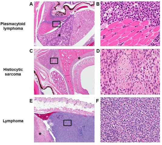
H&E sections of 3 representative hematopoietic tumor types observed in rapamycin treated female mice. Lower magnification (A, C, E. Original magnification 4x) represents an uncommon site of invasion for hematopoietic tumors in C57BL/6 mice. Higher magnification (B, D, F. Original magnification 40x. The region is approximately indicated by the box in A, C, E, respectively) shows the morphology of each cancer cell type. (A) Sheets of neoplastic round cells infiltrate the soft tissues of the head including the Harderian gland (*) and retrobulbar musculature (indicated by box). (B) Neoplastic round cells have a plasmacytoid morphology characterized by an eccentric nucleus, perinuclear halo, and occasionally strongly eosinophilic cytoplasm. (C) A round cell neoplasm morphologically consistent with histiocytic sarcoma in a high dose rapamycin treated female expands and infiltrates the meninges (*) and soft tissues of the head (indicated by box). (D) A round cell neoplasm morphologically consistent with histiocytic sarcoma. Neoplastic histiocytes with abundant eosinophilic cytoplasm and occasional multinucleated giant cells separate and surround nerves. (E) Subcutaneous round cell neoplasm in a high dose rapamycin treated female associated with severe necrosis (*). Box indicates neoplasm composed of sheets of neoplastic lymphocytes. (F) Neoplastic lymphocytes have scant to moderate cytoplasm, marked anisocytosis and anisokaryosis, and frequent mitotic cells.
Figure 3—figure supplement 2. Cancer incidence of male mice with rapamycin injection at 8 mg/kg/day for 3 months.
(A) Hematopoietic cancer incidence of rapamycin-treated (15 male) and vehicle-treated (14 male) mice. (B) Incidence of multiple organ invasion of hematopoietic tumors in rapamycin-treated (9 male) and vehicle-treated (14 male) hematopoietic tumor-bearing mice. (C) Non-systemic hematopoietic cancer incidence of rapamycin-treated (15 male) and vehicle-treated (14 male) mice. **p<0.01.