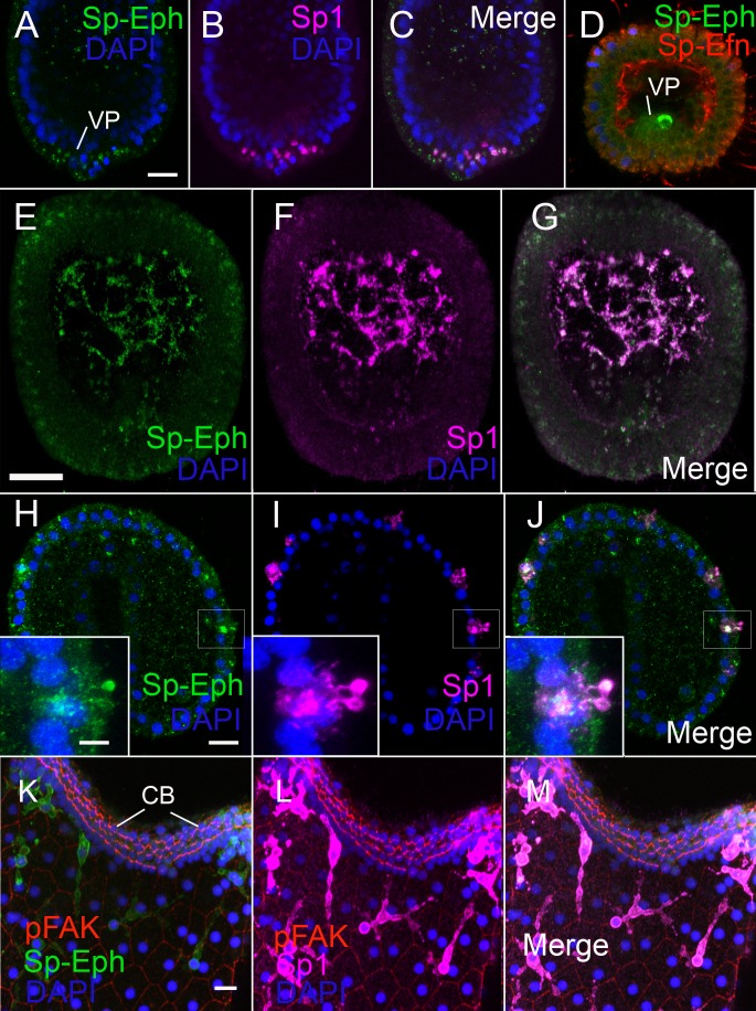
Figure 3. Pigmented immunocytes express Sp-Eph as they differentiate in the vegetal plate and throughout their migration and insertion into ectoderm.
(A–C) In blastulae, SpEph can be detected in cells beginning to express the pigmented immunocyte marker Sp1. The cells are in the vegetal plate and immunoreactivity is strongest in foci adjacent to or overlapping with foci of Sp1 immunoreactivity (MeOH fixation). (D) At stages in which cells expressing Sp-Eph are releasing from the vegetal plate, Sp-Efn can be detected on process on the basal surfaces of ectodermal cells. Here a single pigmented immunocyte progenitor is emerging from the vegetal plate (PEM fixation). (E–G) Pigmented immunocyte precursors expressing surface Sp1 in the blastocoel also express Sp-Eph (MeOH fixation). (H–J) In gastrulae, pigmented immunocytes expressing Sp-Eph have inserted into the ectoderm. Inset images indicate that processes of immunocytes that extend through the ectoderm express Sp-Eph (MeOH fixation). See supplemental data Video 5. (K–M). In early larvae immunocytes within the ectoderm continue to express Sp-Eph on their surface (MeOH fixation). Bars = 15 µm

