. 2016 Aug 23;5:e13424. doi: 10.7554/eLife.13424

Figure 3. Increased cofilin activity in the hippocampus mediates the spine loss associated with sleep deprivation.

(A) Five hours of sleep deprivation leads to a reduction in cofilin phosphorylation at serine 3 in the hippocampus. A representative blot is shown. Each band represents an individual animal. (n = 13–14, Student’s t-test p=0.0090). (B) Mice were injected with pAAV9-CaMKIIα0.4-eGFP or pAAV9-CaMKIIα0.4-cofilinS3D-HA into the hippocampus to drive expression of eGFP or the mutant inactive form of cofilin (cofilinS3D) in excitatory neurons. This inactive mutant form of cofilin was made by substituting serine 3 for aspartic acid, which mimics a phosphoserine residue. An HA-tag was included to discriminate between mutant and endogenous cofilin. (C) A representative image showing that viral eGFP expression was restricted to the hippocampus. (DF) CofilinS3D expression was excluded from astrocytes in area CA1 as indicated by a lack of co-labeling (F) between viral cofilin (D) and GFAP expression (E). Scale bar, 100 µM. (G) Virally delivered cofilinS3D protein levels were approximately 75% (blue bar) of wild-type cofilin levels (green bar). Wild-type cofilin levels were not significantly affected by expression of cofilinS3D. An HA-tag antibody was used to detect the mutant inactive form of cofilin. (n = 4). (H) Hippocampal cofilinS3D expression prevents spine loss in apical/basal dendrites of CA1 neurons that is associated with sleep deprivation (n = 6, Student’s t-test, p>0.05). (I) Hippocampal cofilinS3D expression prevents the decrease in apical/basal dendritic spine length in neurons of hippocampal that is caused by sleep deprivation (n = 6, Student’s t-test, p>0.05). (J) Sleep deprivation does not alter the number of any spine type in apical/basal dendrites of CA1 neurons in the hippocampus of mice expressing cofilinS3D (n = 6, Student’s t-test, p>0.05). (K) Sleep deprivation does not attenuate apical/basal spine density at any distance from the soma in mice expressing cofilinS3D (n = 6, Student’s t-test, p>0.05). NSD: non-sleep deprived, SD: sleep deprived. Values represent the mean ± SEM. **p=0.0090. Student’s t test. See also Figure 3—figure supplement 1. For separate analyses of apical and basal spine numbers see Figure 3—figure supplement 2.

DOI: http://dx.doi.org/10.7554/eLife.13424.008

Figure 3—source data 1. Sleep deprivation reduces cofilin phosphorylation in the hippocampus.
The data source file contains the relative optical density values (in arbitrary units) of the pcofilin and cofilin western blots for each individual animal of the non-sleep deprived (NSD) and sleep deprived (SD) group.
DOI: 10.7554/eLife.13424.009

Figure 3.

Figure 3—figure supplement 1. Sleep deprivation does not alter cofilin phosphorylation in the prefrontal cortex.

Figure 3—figure supplement 1.

Five hours of sleep deprivation does not lead to a reduction in cofilin phosphorylation at serine 3 in the prefrontal cortex. A representative blot is shown. Each band represents an individual animal. (Student’s t test p>0.5).
Figure 3—figure supplement 1—source data 1. Sleep deprivation does not alter cofilin phosphorylation in the prefrontal cortex.
The data source file contains the optical density values (in arbitrary units) of the pcofilin and cofilin western blots for each individual animal of the non-sleep deprived (NSD) and sleep deprived (SD) group.
DOI: 10.7554/eLife.13424.011
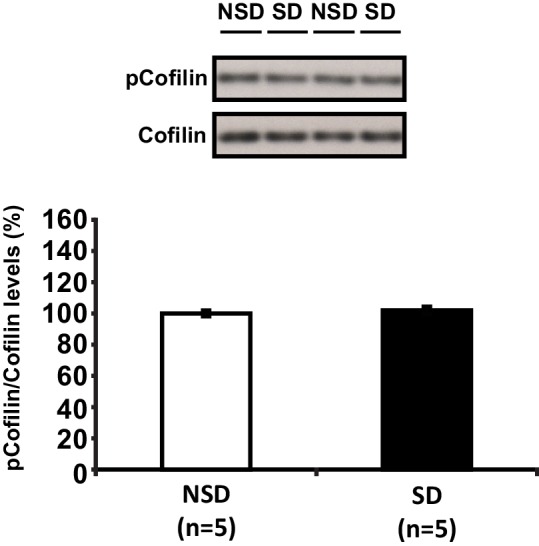
Figure 3—figure supplement 2. CofilinS3D expression prevents sleep deprivation-induced reductions in spine numbers and dendrite length in both basal and apical dendrites of CA1 neurons.

Figure 3—figure supplement 2.

(A) In mice expressing cofilinS3D, sleep deprivation does not decrease the spine density of basal and apical dendrite of CA1 neurons (6 animals per group, 4 neurons per animal). (B) In mice expressing cofilinS3D, sleep deprivation does not cause a decrease in the length of basal and apical dendrite length of CA1 neurons (6 animals per group, 4 neurons per animal). (C) In mice expressing cofilinS3D, sleep deprivation does not cause a reduction in the total number of spines for each subtype in basal dendrites of CA1 neurons with exception of the branched spines (6 animals per group, 4 neurons per animal, Student’s t test p>0.5, branched spines Student’s t test p<0.05). (D) In mice expressing cofilinS3D, sleep deprivation does not cause a decrease in the total number of spines for each subtype in apical dendrites of CA1 neurons with exception of the branched spines (6 animals per group, 4 neurons per animal, Student’s t test p>0.5, branched spines Student’s t test p<0.05). (E) In mice expressing cofilinS3D, sleep deprivation does not reduce the spine density of basal dendrites at any distance from the soma (6 animals per group, 4 neurons per animal, Student’s t test p>0.05). (F) In mice expressing cofilinS3D, sleep deprivation reduces spine density of apical dendrites only at a 180 µm distance from the soma (6 animals per group, 4 neurons per animal, Student’s t test p>0.05). (G) In mice expressing cofilinS3D, sleep deprivation does not decrease the spine density in any dendritic branch of the basal dendrites of CA1 neurons (6 animals per group, 4 neurons per animal, Student’s t test p>0.05). (H) In mice expressing cofilinS3D, sleep deprivation does not reduce the spine density in any dendritic branch of apical dendrites of hippocampal CA1 neurons (6 animals per group, 4 neurons per animal, Student’s t test p>0.05). NSD: non-sleep deprived, SD: sleep deprived. Values represent the mean ± SEM. *p<0.05 by Student’s t test.