. 2016 Aug 23;5:e13424. doi: 10.7554/eLife.13424

Figure 4. Increased cofilin activity in the hippocampus mediates the memory and synaptic plasticity deficits associated with sleep deprivation.

(A) Mice expressing eGFP or cofilinS3D were trained in the hippocampus-dependent object-place recognition task. Half of the groups were sleep deprived for 5 hr and all mice were tested 24 hr later. Hippocampal cofilinS3D expression prevents memory deficits caused by sleep deprivation (n = 9–10, two-way ANOVA, effect of virus F1,35 = 18.567, p=0.0001; effect of sleep deprivation F1,35 = 2.975, p=0.093; interaction effect F1,35 = 4.567, p=0.040; eGFP SD group versus other groups, p<0.05). The dotted line indicates chance performance (33.3%). (B, C) Following 5 hr of sleep deprivation, long-lasting LTP was induced in hippocampal slices by application of four 100 Hz trains, 1 s each, spaced 5 min apart to the Schaffer collateral pathway. Five hours of sleep deprivation impairs long-lasting LTP in slices from mice expressing eGFP (n = 6–7, two-way ANOVA, effect of virus F1,10 = 21.685, p<0.001). In contrast, virally delivered cofilinS3D prevents sleep deprivation-induced deficits (n = 5, two-way ANOVA, effect of virus F1,8 = 0.016, p>0.902). NSD: non-sleep deprived, SD: sleep deprived. Values represent the mean ± SEM. *p<0.05 by posthoc Dunnet’s test, **p<0.01 by Student’s t test. See also Figure 4—figure supplement 1.

DOI: http://dx.doi.org/10.7554/eLife.13424.013

Figure 4—source data 1. CofilinS3D expression prevents memory deficits in the object-location memory task caused by sleep deprivation.
The data source file contains the object exploration times for the displaced (DO) and non-displaced objects (NDO1, NDO2) for all individual animals of each group.
DOI: 10.7554/eLife.13424.014

Figure 4.

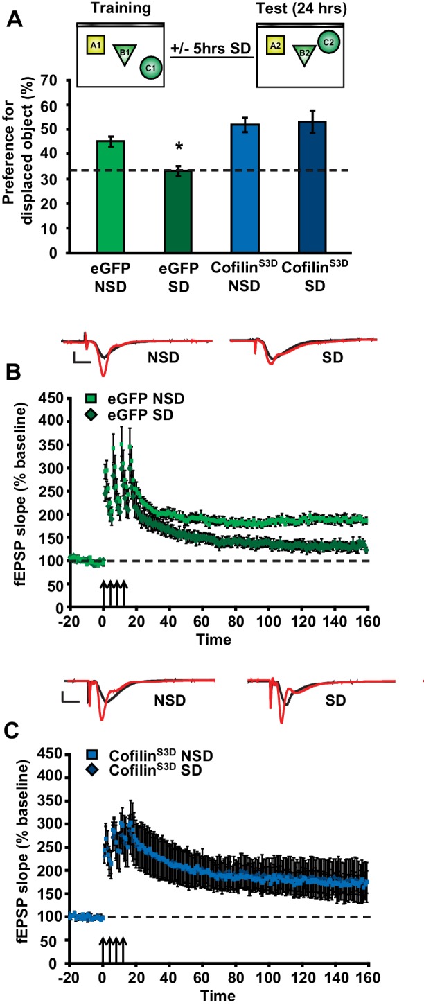
Figure 4—figure supplement 1. CofilinS3D expression in hippocampal neurons does not affect exploratory activity, anxiety levels, or basal synaptic transmission.

Figure 4—figure supplement 1.

(A) Expression of the catalytically inactive cofilinS3D in hippocampal neurons does not affect the total time spent exploring objects during training in the object place recognition task (ANOVA F1,35 = 1.026, p=0.318). All groups show a decrease in the total object exploration time during the training sessions (n = 9–10, two-way ANOVA, effect of session F2,70 = 54.060, p = 0.0001; interaction effect F2,70 = 0.880, p=0.419). (B) Mice expressing cofilinS3D in hippocampal neurons spent a similar amount of time in the enclosed arm of the zero maze as eGFP expressing mice indicating normal anxiety-related behavior (n = 7, Student’s t test, p=0.632). (C) Mice expressing cofilinS3D in hippocampal neurons had a similar number of transitions in the zero maze as eGFP expressing mice indicating normal locomotor activity (n = 7, Student’s t test, p=0.849). (D) CofilinS3D expression did not alter the formation of short-term object location memories measured one hour after training (n = 7–8, Student’s t test, p=0.42). (E, F) Input-output curves relating the amplitude of the presynaptic fiber volley to the initial slope of the corresponding fEPSP at various stimulus intensities are similar in slices from eGFP and cofilinS3D in slices from sleep deprived and non-sleep deprived mice (n = 5, eGFP NSD vs SD group, Student’s t test p=0.75; cofilinS3D, NSD vs SD group, Student’s t test p=0.17). (G, H) Paired-pulse facilitation, a short-term form of synaptic plasticity, was not changed in slices from eGFP and cofilinS3D in slices from sleep deprived and non-sleep deprived mice (n = 5, eGFP NSD vs SD group two-way repeated-measures ANOVA, F1,8= 0.393, p=0.545) (cofilinS3D NSD vs SD group two-way repeated-measures ANOVA, F1,8= 3.056, = 0.114). NSD: non-sleep deprived, SD: sleep deprived. Values represent the mean ± SEM.
Figure 4—figure supplement 1—source data 1. CofilinS3D expression in hippocampal neurons does not affect exploratory activity.
(A) The data source file contains the total object exploration times during the three training sessions for each individual animal of all four groups. (B) The data source file contains the time spent in the closed arms of the zero maze for each individual animal of both groups. (C) The data source file contains the number of transitions in the zero maze for each individual animal of both groups. (D) The data source file contains the object exploration times for the displaced (DO) and non-displaced objects (NDO1, NDO2) for each individual animal of both groups.
DOI: 10.7554/eLife.13424.016
Figure 4—figure supplement 2. CofilinS3A expression in hippocampal neurons attenuates the formation of long-term object-location memories but not long-term potentiation induced by spaced-four train LTP.

Figure 4—figure supplement 2.

(A) Mice expressing eGFP or the catalytically active cofilinS3A in hippocampal neurons were trained in the hippocampus-dependent object-place recognition task. Expression of the cofilinS3A does not affect the total time spent exploring objects during training in the object place recognition task (ANOVA F1,18 = 1.919, p=0.183). Both groups show a decrease in the total object exploration time during the training sessions (n = 10, two-way ANOVA, effect of session F2,36 = 11.696, p=0.0001; interaction effect F2,36 = 1.85, p=0.172). (B) During the test session 24 hr after training, eGFP mice preferentially explored the displaced object indicating that they successfully remembered the previous location of the individual objects. In contrast, mice expressing cofilinS3A explored all objects to a similar extent, indicative of a poor memory for the original object locations (eGFP, 46.9 ± 4.2%; cofilinS3A, 34.9 ± 2.1%; Student’s t test, p=0.025). (C) Input-output curves relating the amplitude of the presynaptic fiber volley to the initial slope of the corresponding fEPSP at various stimulus intensities are similar in slices from eGFP and cofilinS3A in slices from non-sleep deprived mice (eGFP n = 6, cofilinS3A n = 8, Student’s t test p = 0.857). (D) Paired-pulse facilitation, a short-term form of synaptic plasticity, was not changed in slices from eGFP and cofilinS3A in slices from non-sleep deprived mice (n = 5–6, eGFP vs cofilinS3A group two-way repeated-measures ANOVA, F1,12= 0.218, p=0.649). (E) Long-lasting LTP was induced in hippocampal slices by application of four 100 Hz trains, 1 s each, spaced 5 min apart to the Schaffer collateral pathway. Virally delivered CofilinS3A expression did not alter LTP expression (n = 5, two-way ANOVA, effect of virus F1,8 = 1.102, p=0.0325). Dotted line indicates chance level performance. Values represent the mean ± SEM. *p<0.05 by Student’s t test.