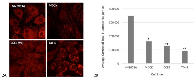
Figure 2.
Figure 2A – Confocal microscopy of Mito Tracker Red CMXRos staining of different cell lines. 2B – Average fluorescence of mitochondrial staining. (* p<0.05, ** p<0.001 compared to NN1003A). Rabbit lens epithelial cell (NN1003A) staining revealed the highest density of mitochondria within the cells compared to canine kidney epithelial cells (MDCK; p<0.05), bovine corneal endothelial cells (CCEE; p<0.001) and human trabecular meshwork cells (TM-5; p<0.001).

