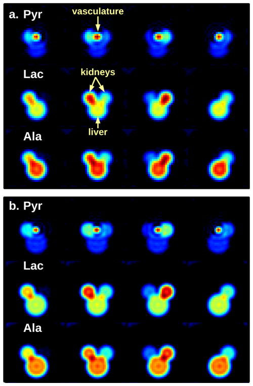
FIG. 2.
Four representative slices from the center of a rat phantom at the 12 s time point. Pyruvate, lactate, and alanine images are shown for (a) parameter set (A) and (b) parameter set (B). The model consists of cylinders representing kidneys, vasculature, and liver/body. Note that only pyruvate is modeled in the vasculature.

