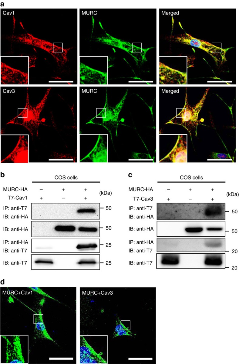
Figure 1. Colocalization of MURC with Cav1 and Cav3 in hPASMCs.
(a) Representative immunostaining images of MURC, Cav1 and Cav3 in hPASMCs. Scale bar, 50 μm. (b,c) COS cells were transfected with pcDNA3.1-hMURC-HA and/or pcDNA3.1-T7-hCav1 (b) or pcDNA3.1-T7-hCav3 (c), and the cell lysates were immunoprecipitated with anti-HA and anti-T7 antibodies. (d) The in situ association of proteins in hPASMCs was assessed by the BiFC assay, which detects fluorescent signals in living cells when proteins associate with each other. Upper, hPASMCs were transfected with phmKGC-MC-hMURC and phmKGN-MC-hCav1. Lower, hPASMCs were transfected with phmKGN-MN-hMURC and phmKGC-MN-hCav3. Scale bar, 50 μm. Uncropped images of blots are shown in Supplementary Fig. 6.

