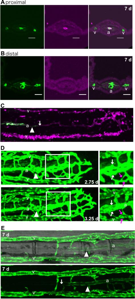
Fig. 2.
Artery and vein separation occurred by rearrangement of the vascular plexus.
(A and B) Frozen cross-sections of different regions of the same fin ray in a Tg(fli1a:EGFP) caudal fin at 7 dpa. The arterial vessel in the regenerate close to the stump (proximal) was associated with smooth muscle cells (red, A). In the distal region of the fin ray (B), the vasculature (green) had separated into 3 vessels, but smooth muscle cells (red) had not yet been recruited. As the fin rays in the regenerate were usually thicker than those in the mature fin rays, they could be easily distinguished. Scale bars, 20 μm. (C) Simultaneous imaging of Tg(kdrl:EGFP) (green) and the trajectory of the blood cells (red) in living Tg(kdrl:EGFP); Tg(gata1a:dsRed) fish. The white arrow points to the distal extent of Tg(kdrl:EGFP) -positive cells. In the more distal side of the regenerate, Tg(kdrl:EGFP) -positive cells were not observed, although the blood flow showed 3 distinct streams, presumed to be the future artery and veins. (D) Representative image of remodeling of the vascular plexus. Time-lapse images of a Tg(fli1a:EGFP) regenerating fin ray at 2.75 and 3.25 dpa. Right panels are enlarged figures of the boxed regions in the left ones. Vascular fusion occurred close to the future artery (white arrows) and future veins (red arrows). (E) A representative regenerating fin ray of a Tg(flt4:YFP) at 7 dpa. A fluorescence image (bottom) and a same image merged with a bright-field image (top) are shown. In the stump, the artery did not have Tg(flt4: YFP) -positive cells except in a small region close to the artery in the regenerate. On the other hand, the artery in the regenerated region was Tg(flt4:YFP) positive. The amputation site is indicated by the white arrowhead. a, artery; v, vein.

