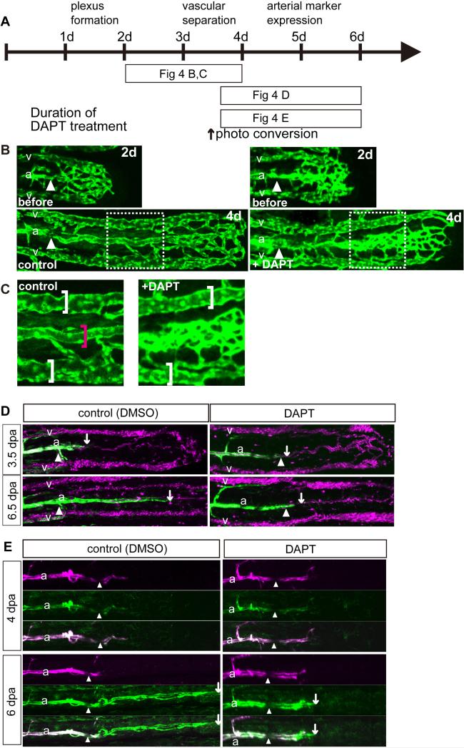
Fig. 4.
Notch signaling was essential for vascular separation and arterial differentiation during fin regeneration. (B-D) Zebrafish caudal fins were clipped and then treated with the γ-secretase inhibitor DAPT or control DMSO as indicated in “A.” (B) Inhibition of Notch signaling caused vascular separation defects. DAPT treatment from 2 dpa (before ; 2d) to 4 dpa (+ DAPT; 4d) caused vascular separation defects in the artery but not in the veins. “C” is an enlarged image of the boxed region in “B.” Inhibition of Notch signaling from 3.5 to 6.5 dpa inhibited a transgene expression from kdrl promoter, which represents arterial gene expression (D). Tg(kdrl:EGFP); Tg(gata1a:dsRed) fish were treated with DAPT from 3.5 to 6.5 dpa. White arrows point to the distal end of the area of Tg(kdrl:EGFP) positive cells. The blood flow had already separated into 3 vessels at 6 dpa in both the control and some fin rays of the DAPT-treated fish. However, Tg(kdrl:EGFP) -positive cells were not observed in the DAPT-treated fish. (E) Inhibition of Notch signaling in a Tg(kdrl:Kaede) fish from 4 to 6 dpa inhibited arterial gene expression. Kaede-expressing arterial cells were labeled (red) by photo-conversion at 4 dpa. Soon after conversion, the fish were treated with DAPT up to 6 dpa. The amputation site is indicated by the white arrowheads in all photos. White arrows point to the the distal end of the area of Tg(kdrl:Kaede) positive cells.

