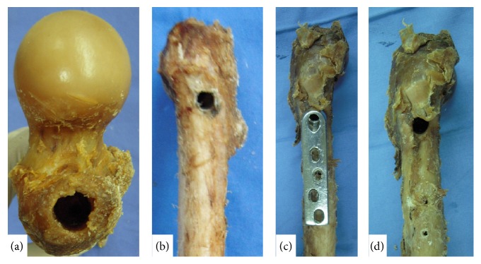
Figure 1.
(a) An opening was created on the tip of the greater trochanter following removal of the PFNA-II (16.5 mm). (b) An opening was created on the lateral wall of the proximal femur following removal of the PFNA-II. (c) DHS position on the proximal femur. (d) An opening was created on the lateral wall of the proximal femur following removal of the DHS (12.5 mm).

