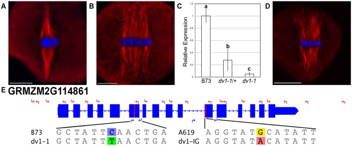
Figure 1.
Two alleles of dv1 show a divergent spindle phenotype. Meiocytes were stained using immunofluorescence with a primary antibody specific to α-tubulin. Scale bars for each image represent 10 μm. (A) Wild type spindle showing highly focused spindle poles; (B) dv1-1/dv1-1 spindle showing splayed, divergent poles; (C) Expression of the dv1-1 allele of ZmKin6 adjusted to the B73 wild type allele. Error bars represent 95% confidence intervals, groups of significant difference are designated with lowercase letters; (D) dv1-1/dv1-IG heteroallelic mutant is similar to the dv1-1 homozygote; (E) Gene model of ZmKin6 highlighting the location of the two dv1 alleles, the dv1-1 stop codon in the sixth exon and the dv1-IG transversion in the motor domain. Coordinates along chromosome 2 are shown above. The locations of sequencing primers are shown with red arrows while the locations of genotyping primers are shown with blue arrows. The reference and mutant sequences of each allele are shown below the gene diagram.

