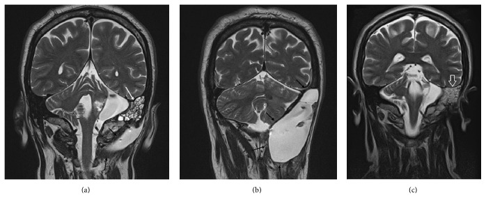
Figure 2.
(a) Coronal T2-weighed image. Hyperintense CSF leak to the posterior skull base (black arrows). CSF leak in mastoid cells (white arrow). (b) Coronal T2-weighed image. Massive hyperintense CSF leak extended between the latera skull base and the fist cervical vertebra. Cerebellum compression visible (black arrows). (c) Postoperative coronal T2-weighed image. No CSF leak. Hyperintense signal to the mastoid air cells (white open arrow).

