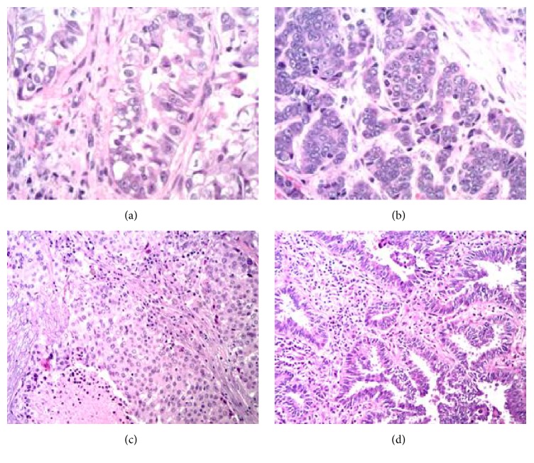
Figure 2.
H&E stain of two specimens is shown. Panel (a) shows the glandular tumor component from specimen 1. Panel (b) shows the neuroendocrine tumor component from specimen 1. Panel (c) shows an invasive adenocarcinoma, solid predominant, poorly differentiated tumor, from specimen 2. Panel (d) shows the minimally invasive adenocarcinoma, nonmucinous type, from specimen 2.

