Figure 3.
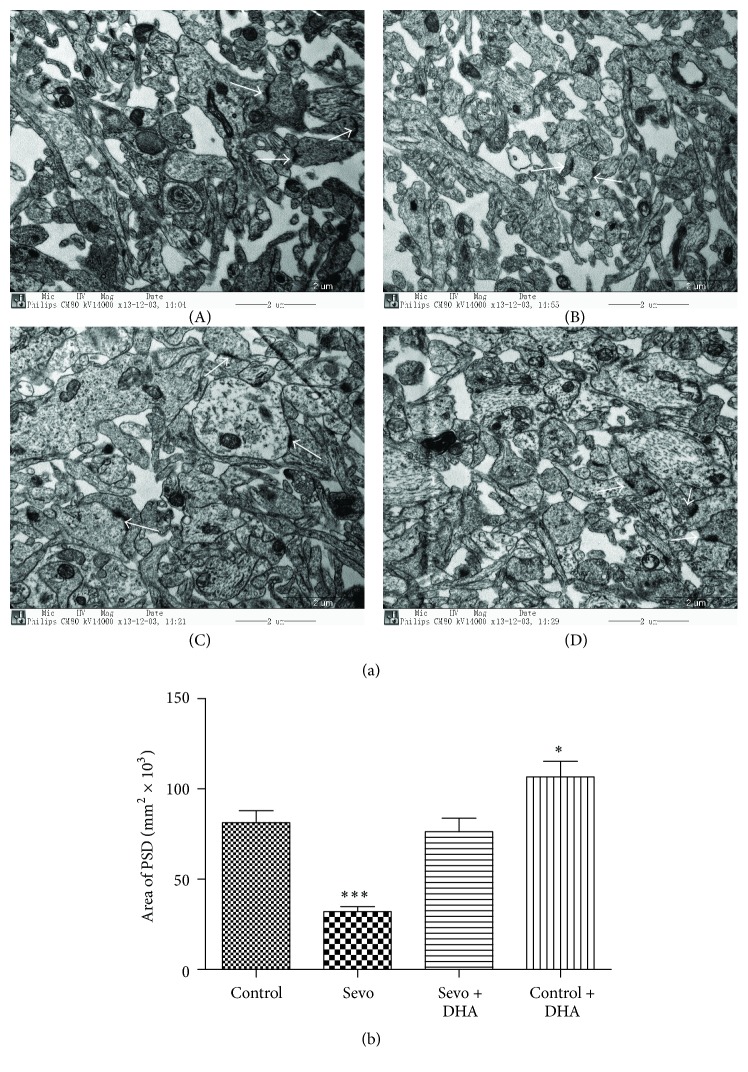
DHA alleviated synaptic ultrastructure impairment after sevoflurane exposure. (a) Hippocampal synaptic ultrastructure in the control group ((a)(A)), Sevo group ((a)(B)), Sevo + DHA group ((a)(C)), and control + DHA group ((a)(D)) was analyzed by transmission electron microscopy. White arrows showed postsynaptic densities (PSD). Scale bar = 2 μm. (b) Quantitation of PSD areas was represented. Data are shown as mean ± SEM; ∗∗∗ P < 0.001 and ∗ P < 0.05 comparing with control group, n = 9 for each group. Sevo = sevoflurane.

