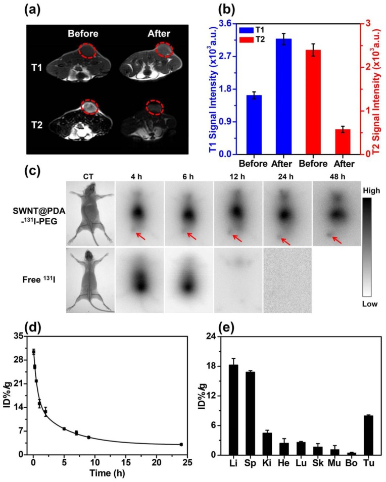
Figure 4.
In vivo behaviors of SWNT@PDA-131I-PEG. (a) In vivo T1- (upper) and T2-weighted MR images (bottom) of a 4T1 tumor-bearing mouse taken before injection (left) and 24 h post injection (right) of SWNT@PDA-PEG/Mn. Obvious brightening and darkening effects showed up in the tumor after i.v. injection of SWNT@PDA-PEG/Mn by T1- and T2-weighted MR imaging, respectively. (b) T1- (left) and T2-weighted (right) MR signals intensities of the tumor before and 24 h post i.v. injection of SWNT@PDA-PEG/Mn, which offered strong tumor contrasts under both T1- and T2-weighted MR imaging modes. (c) Gamma imaging of SWNT@PDA-131I-PEG-treated mice and free 131I-treated mice. Notably, free 131I was completely excreted after 12 h, while SWNT@PDA-131I-PEG showed obvious tumor accumulation. (d) The blood circulation of SWNT@PDA-131I-PEG after i.v. injection determined by gamma-counting. (e) The biodistribution of SWNT@PDA-131I-PEG in 4T1 tumor-bearing mice measured at 24 h p.i. Li: liver; Sp: spleen; Ki: kidney; He: heart; Lu: lung; Sk: skin; Mu: muscle; Bo: bone; Tu: tumor.

