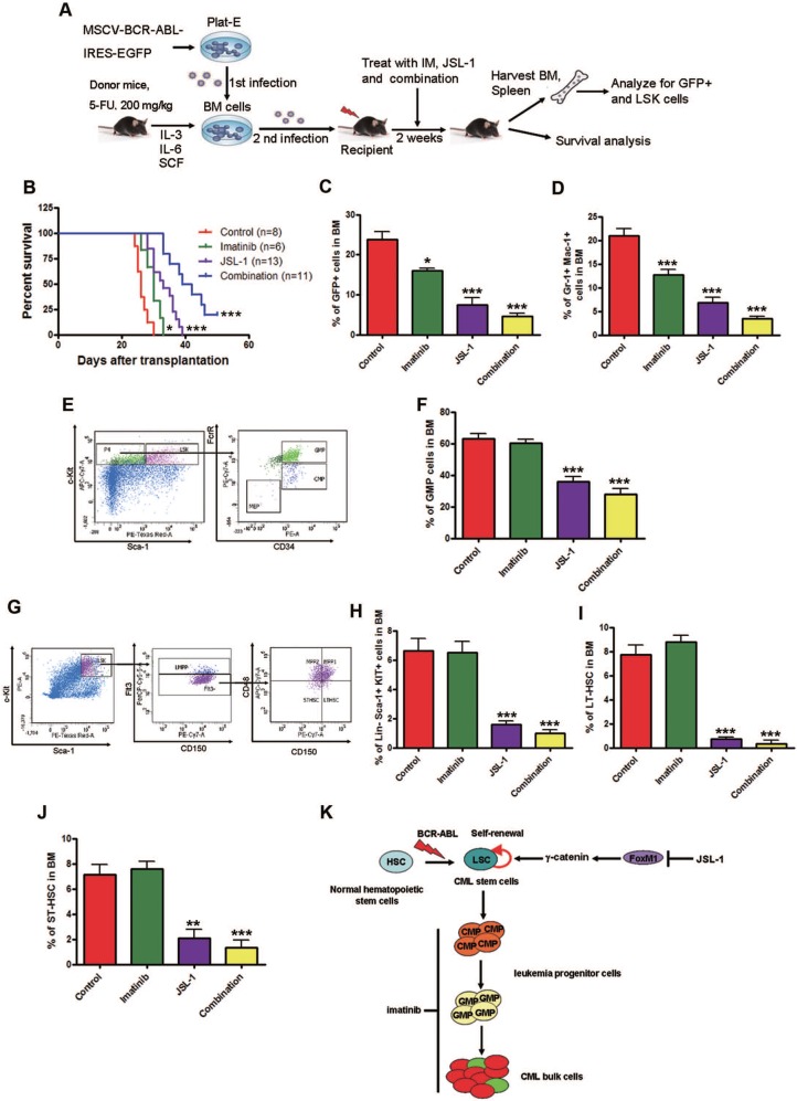
Figure 7.
Pharmacological inhibition of γ-catenin reduces growth of CML LSCs in mice. (A) A schematic strategy to generate the retroviral BCR-ABL-driven CML mouse model and drug treatment. (B) JSL-1 treatment alone or in combination with imatinib significantly prolonged survival of CML mice. Kaplan-Meier survival curves were shown, * P=0.0178, *** P<0.0001, log-rank test. (C) GFP+ cells in BM. (D) GFP+ myeloid cells (Gr-1+ Mac-1+) cells in BM. (E) Representative flow cytometry plots GMP in BM of mice treated with imatinib and JSL-1. (F) GMP cells in BM. (G-J) Analysis of LSCs in the BM cells of CML mice received treatment with imatinib ± JSL-1. Control (n=13), imatinib (n=10), JSL-1 (n=5), and combination (n=5). (G) Representative flow cytometry plots of LSK cells, LT-HSCs and ST-HSCs in BM of mice treated with imatinib and JSL-1. (H-J) Results for the GFP+ population in BM: LSK cells (H), LT-HSCs (I), and ST-HSCs (J). * P<0.05, ** P<0.01, *** P<0.0001. (K) A proposed model of HDACi JSL-1 action against self-renewal of CML LSCs. Quiescent CML LSCs are insensitive to imatinib. γ-Catenin is required for survival and self-renewal of LSCs. Loss of γ-catenin in combination with imatinib eliminates LSCs in CML.

